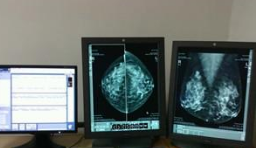
An international research team pioneered a new type of X-ray mammography that captures a three-dimensional X-ray image of the breast at a dose that is about 25 times lower than the two-dimensional radiography currently used. At the same time, the new method can also increase the spatial resolution of the generated 3D high-energy X-ray computed tomography (CT) diagnostic image by 2 to 3 times. Related research papers were published on the same day in the online edition of the Proceedings of the National Academy of Sciences.
The currently used breast cancer scanning technique is "Double View Digital Mammography", which has the drawback of providing only two images of breast tissue, which explains why 10% to 20% of breast tumors cannot be detected. In addition, this kind of photography occasionally has abnormalities that cause misdiagnosis of breast cancer.

While CT, an X-ray technique, can generate accurate three-dimensional visual images of human organs, it cannot be used frequently in the diagnosis of breast cancer because it may cause long-term effects on radiation-sensitive organs such as breasts. The risk is too high.
New technologies are expected to overcome the above limitations. Researchers are currently testing this technology with synchrotron X-rays, which, once put into use in hospitals, will make CT scanning one of the diagnostic tools that complement dual-view digital mammography.
The use of high-energy X-ray and phase contrast imaging technology, coupled with complex new EST mathematical algorithms, enables the reconstruction of CT images based on X-ray data, making CT scans promising for early breast cancer screening and a powerful tool for combating breast cancer. The body tissue will become more transparent under the illumination of high-energy X-rays, so the required radiation dose can be significantly reduced by about 6 times. Phase contrast imaging also allows for fewer X-rays when shooting the same photo, and the EST algorithm also achieves the same image quality with 4x less radiation. In this way, the research team took 512 breast images from a number of different angles and formed a three-dimensional image with higher definition, contrast and overall image quality than traditional mammography.
According to researchers, these high-quality, high-energy X-ray CT images are the result of 10 years of struggle for the European Synchrotron Radiation Source (ESRF) Research Center, as well as the University of Munich and the University of California, Los Angeles. They also said that the next research goal is to achieve early visualization of other human diseases based on this technology, and to develop a suitable X-ray source in an effort to realize the clinical application of the technology as soon as possible.
Multiple Beam Angles street lights,LED Street light,Led White Light Street lamp
Kindwin Technology (H.K.) Limited , https://www.ktlleds.com